Nerve Synapses Close Up
Section titled “Nerve Synapses Close Up”Return to February 2006 Newsletter
Here is an actual photograph of the terminal of an incoming cell on the top, a tiny part of the receiving cell on the bottom, and the synapse (here labeled synaptic cleft) in between.
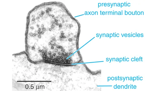
The top part (presynaptic axon terminal bouton) is the continuation of a tiny output fiber from a nerve cell that, in the scale of this photograph, could be as far as thirty miles away. The postsynaptic dendrite at the bottom is just a tiny part of a receiving cell, like the one illustrated with all the green and red dots. This is a close-up of one of the green or red dots.
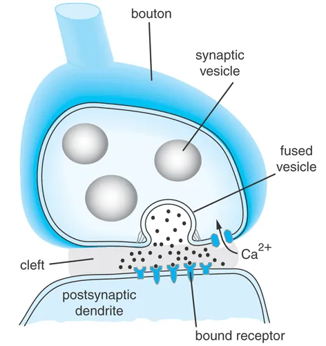
The synaptic cleft is the gap between cells. The tiny pouches called synaptic vesicles contain a neurotransmitting chemical such as serotonin, which will diffuse across the gap. This neurotransmitting chemical will then signal the receiving nerve cell to fire or to refrain from firing.
You've probably heard of "selective serotonin-reuptake inhibitors," which are antidepressant drugs such as Prozac. An SSRI acts at the synapse. After the serotonin is released into the synaptic cleft, the sending cell membrane usually pumps the serotonin back out of the cleft, to re-use it for the next signal. Prozac and other SSRIs prevent the cell membrane from scavenging the serotonin back out of the cleft. With more serotonin left in that cleft, there is a better chance of sendinga "happy" signal to the next neuron
Here is a schematic view of a neuron signaling another at the synapse.