The patient came into my office in misery. He'd gone skiing and had awakened the next morning with severe stabbing pains in his back and pelvis. Another doctor had ordered a CT scan. Coming to me for a second opinion, the patient brought the scan on a disk clasped between his fingers. The radiologist's report read "degenerative disk disease, most notably at L1-L2 and L2-L3 with disk space narrowing. Air vacuum signs are seen at L1-L2, L2-L3, and L3-L4. Mild retrolisthesis of L2 on L3 is present." Yikes!
Take a look at the scan and I'll translate that for you.
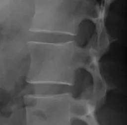
The skier's X-ray
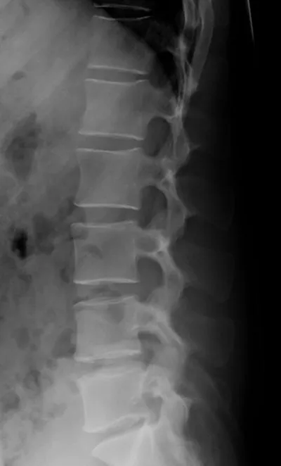
An X-ray showing more normal intervertebral spaces.
On the scan, the block-like things are the vertebral bodies and the triangular/trapezoidal shapes are the vertebral spines, those bumps that run down the middle of your own back. (These are bright white on the patient, more grayed out in the normal spine on the right.) You can see that the fourth vertebral body from the top (the second lumbar, or L-2) has slid backwards relative to the vertebral body below, which is called retrolisthesis. The space between the vertebral bodies in the middle of the image does not show the normal cushioning. The black areas are air bubbles between the vertebral bodies, caused by excessive stress pulling the vertebral bodies apart. There are also bony spurs sticking out from the tops and bottoms of the vertebral bodies in the middle of the image.
When Pictures Deceive
Section titled “When Pictures Deceive”What's a person to think about these changes to the spine? No more skiing? A life of pain?
I told him that I had cared for plenty of people with CT scans just as bad or worse than his. Most of those patients now felt fine. The skier asked if chiropractic could give some immediate relief. I agreed as long as the chiropractor used gentle techniques. I also prescribed potent pain medication. He accepted this recommendation and, sure enough, a week later he felt considerably better. I gave him an exercise book for the low back. He did the exercises every day and within a month his back didn't hurt anymore. Every day, as certainly as he brushes his teeth, he gets down on the floor and does exercises to strengthen the muscles that support his spine.
He told me that his back had actually hurt for years when he drove his car for too long, and he had attributed it to the car seat. Now he could drive his car without pain.
Diagnosis Can Make You Ill
Section titled “Diagnosis Can Make You Ill”Frequently I run into people who tell me with some pride that they have not seen a doctor in many years and that is the reason for their good health. Sadly, in some cases, I must agree.
I'm not talking about side effects from pharmaceuticals or infections picked up in the hospital. I'm talking about a diagnosis that's worse than the condition. I'm talking about planting the idea in your mind that your condition is worse than is actually the case. The advent of imaging studies has exacerbated the problem, because it suggests there is bony damage that cannot be fixed.
Someone goes to the doctor with back pain, gets an imaging study they most likely don't need, then are shown the wear and tear in their joints and bones. The doctor often gives it a label, such as degenerative disk disease. The individual, not surprisingly, concludes they can't get well.
Most often, that is just not the case. They can feel a lot better. They can live a relatively pain-free life.
Pain in the Hip
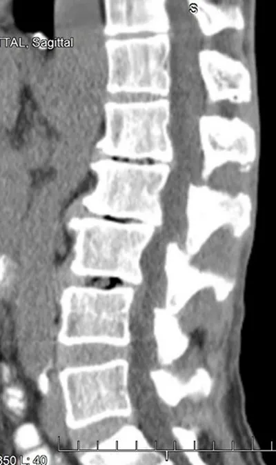
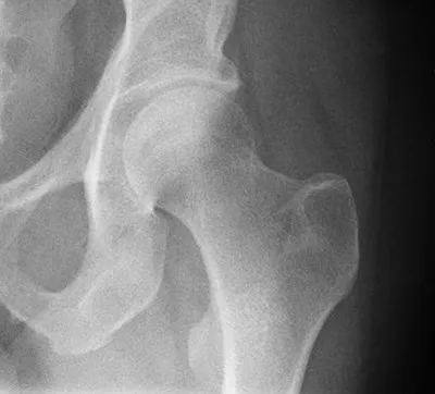
Section titled “Pain in the Hip”To give another example, here's the CT of a heavy equipment operator's hip joint.
The heavy equipment operator
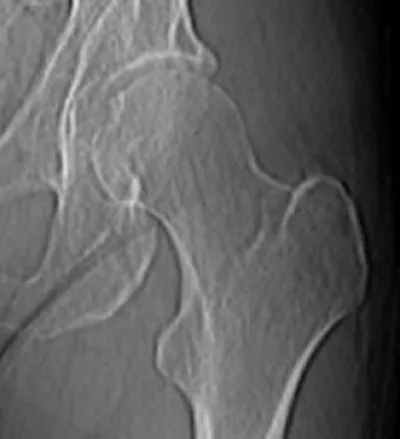
A normal hip joint (Wikipedia)
On the upper left is the pelvic bone with its socket-style hip joint. Rising from the lower right is the top of the femur with its ball fitting into the socket.
The radiologist read this as irregular contour of the acetabular (hip) joint, with mild to moderate osteoarthritis.
In English, this means that both the ball at the top of the femur and the socket in the pelvic joint, which should have smooth surfaces, do not.
OK, his hip joint was not perfect, but I've seen worse. However my patient's pain was rendering him unable to complete his work responsibilities. In fact he was having pain so severe it would drop him to the floor without warning. In addition, he was self-employed and unable to work, so he took a significant financial hit.
While opiates are a curse for many people, they can be a blessing for others and they were a blessing for him for a few days while the acute pain subsided. Once we had achieved that, I sent him to a physical therapist. She looked at the image above and told him that she was not certain that the physical therapy would be all he needed. She left open the option of surgery should her methods not suffice. He too, like the skier, was of a compulsive nature and did the exercises she taught him, as regularly as a monk goes to prayer.
Within two weeks he was back on his excavating equipment and within six weeks was out in the mountains hiking, as was his habit. The last I heard, he'd ascended the steep, rugged trail up Mount Tyler with no difficulty or pain whatever.
Most People Can Get Better
Section titled “Most People Can Get Better”These are two heartening stories. With physical therapy, religiously applied, successes are the most common outcomes. But all too often people see me with chronic inadequately treated pain, telling me that their x-rays show bone spurs, disk narrowing, and other abnormalities. They have been told that these anatomic problems are causing their pain. The implication is that unless the bones are somehow changed they will always be in pain. This diagnosis leaves them in a dark night of disability with no awareness that there could be a dawn.
Our Marvelous Bodies
Section titled “Our Marvelous Bodies”Our musculoskeletal frame is composed not just of bones and joints. As important are the muscles, ligaments and tendons integrated with it. The nervous system carries signals to and from these joints. The central nervous system composes the pain signal, a signal which varies depending upon our cognitive understanding and our emotional state. A CT scan or MRI shows us more than ever we could see before, but still just a tiny fraction of the situation.
Whether the muscles are strong enough or limber enough to make everything work well, whether all these complex systems are normal or not – that is the realm of specially trained physicians and physical therapists. When we see an x-ray or CT scan and we see the bones, we see but a fraction of what we are.
You Are More Than The Sum Of Your Parts
Section titled “You Are More Than The Sum Of Your Parts”Anatomy is not destiny. The MRI and the CT scan can tell us a great deal that is helpful, but they can also be a hindrance, making a person feel limited and disabled.
You may have crow's feet and laugh lines, but that does not mean you are not beautiful. You may have imperfect images on your CT scan, but that does not mean you cannot dance.